

Genes, ethics and society
View Sequence overviewStudents will:
- identify the backbone and bases that form the structure of a DNA molecule.
- identify complementary bases in a DNA molecule.
- construct a model of a DNA molecule.
Students will represent their understanding as they:
- construct a model of a DNA molecule.
- draw a labelled diagram of a model of DNA.
- use the model to predict the impact of damage to DNA.
Assessment is formative.
Feedback might focus on students’ ability to:
- construct a model of a DNA molecule.
- identify the advantages and disadvantages of the DNA model.
- describe the impact of damaged DNA.
Potential summative task
Students working at the achievement standard should:
- select and construct appropriate representations, including models, to organise and process data and information.
Whole class
Genes, ethics and society Slides
Each group
4 x pipe cleaners
4 x paper straws in different colours
Scissors
Each student
Modelling DNA Resource sheet
Student notebook
Lesson
Re-orient
Discuss the analogies from the previous lesson, including the wool chromosome, the gold chain and students’ own analogies.
Discuss the relationship between DNA, chromosomes and genes.
The Inquire phase allows students to cycle progressively and with increasing complexity through the key science ideas related to the core concepts. Each Inquire cycle is divided into three teaching and learning routines that allow students to systematically build their knowledge and skills in science and incorporate this into their current understanding of the world.
When designing a teaching sequence, it is important to consider the knowledge and skills that students will need in the final Act phase. Consider what the students already know and identify the steps that need to be taken to reach the level required. How could you facilitate students’ understanding at each step? What investigations could be designed to build the skills at each step?
Read more about using the LIA FrameworkIdentifying and constructing questions is the creative driver of the inquiry process. It allows students to explore what they know and how they know it. During the Inquire phase of the LIA Framework, the Question routine allows for past activities to be reviewed and to set the scene for the investigation that students will undertake. The use of effective questioning techniques can influence students’ view and interpretation of upcoming content, open them to exploration and link to their current interests and science capital.
When designing a teaching sequence, it is important to spend some time considering the mindset of students at the start of each Inquire phase. What do you want students to be thinking about, what do they already know and what is the best way for them to approach the task? What might tap into their curiosity?
Read more about using the LIA FrameworkWhat do we know?
Pose the question: What do you know about DNA?
Brainstorm where students have heard of DNA previously, what pictures they may have seen of DNA, and what its function may be.
- What images or symbols come to mind when you think of DNA?
- Have you ever seen the double helix shape before? Where?
- What questions do you have about DNA?
- What words do you associate with DNA?
✎ STUDENT NOTES: Brainstorm what you know about DNA structure and any questions that may arise.
Pose the question: What does DNA look like?
The Inquire phase allows students to cycle progressively and with increasing complexity through the key science ideas related to the core concepts. Each Inquire cycle is divided into three teaching and learning routines that allow students to systematically build their knowledge and skills in science and incorporate this into their current understanding of the world.
When designing a teaching sequence, it is important to consider the knowledge and skills that students will need in the final Act phase. Consider what the students already know and identify the steps that need to be taken to reach the level required. How could you facilitate students’ understanding at each step? What investigations could be designed to build the skills at each step?
Read more about using the LIA FrameworkThe Investigate routine provides students with an opportunity to explore the key ideas of science, to plan and conduct an investigation, and to gather and record data. The investigations are designed to systematically develop content knowledge and skills through increasingly complex processes of structured inquiry, guided inquiry and open inquiry approaches. Students are encouraged to process data to identify trends and patterns and link them to the real-world context of the teaching sequence.
When designing a teaching sequence, consider the diagnostic assessment (Launch phase) that identified the alternative conceptions that students held. Are there activities that challenge these ideas and provide openings for discussion? What content knowledge and skills do students need to be able to complete the final (Act phase) task? How could you systematically build these through the investigation routines? Are there opportunities to build students’ understanding and skills in the science inquiry processes through the successive investigations?
Read more about using the LIA FrameworkMaking DNA models
Remind students of the difference between an analogy (something similar that helps understanding) and a model (used to represent, for calculation and prediction).
(Slide 20) Compare the images of a ladder and DNA. Identify the strengths and limitations of the analogy:
- Both have sides that are connected by rungs in the centre.
- DNA is much smaller and twisted.
(Slide 21) Invite students to build a model of DNA that could be used to predict how changes may affect its function.
Provide students with a copy of Modelling DNA Resource sheet and the pipe cleaners, paper straws and scissors.
Guide students to construct their DNA model.

- Cut the paper straws into 2 cm long sections. Each coloured straw section represents a particular DNA base. Record the colours you will use for each base in your model DNA.
- Cut two of the pipe cleaners into 6 cm long segments. These will form the “rungs” on the DNA ladder.
- Thread matching pairs of DNA bases (adenine paired with thymine, cytosine paired with guanine) onto each pipe cleaner segment.
- Lay the remaining two pipe cleaners on the table in parallel, approximately 4 cm apart. This will form the sides of the DNA ladder (the sugar-phosphate backbone of the DNA).
- Place the matched pairs of DNA bases across the sides of the ladder (sugar-phosphate backbone) so that they form the rungs of the ladder. Check that they are equal distances apart from each other.
- Connect the pipe cleaner rungs (DNA bases) to the sides of the DNA ladder (sugar-phosphate backbone).
- Gently twist the sides of the DNA ladder so that it forms a double helix.
✎ STUDENT NOTES: Draw a labelled diagram of the DNA model.
DNA mutations and cystic fibrosis
DNA provides the genetic instructions needed to build proteins.

DNA is the molecule that carries the instructions for how living things grow, function, and reproduce. It is often called the “genetic code” of life. The structure of DNA looks like a twisted ladder (an analogy) and is known as a double helix:
- The sides of the ladder are called the “DNA backbone” and are made from sugar and phosphate molecules.
- The rungs of the ladder are made from pairs of chemical DNA bases.
There are four bases in DNA:
- Adenine (A)
- Thymine (T)
- Cytosine (C)
- Guanine (G)
The DNA bases always pair in the same way:
- A pairs with T
- C pairs with G
DNA provides the genetic instructions needed to build proteins. Proteins are essential molecules that perform many functions within living organisms, including forming body structures, acting as enzymes, transporting substances, and regulating chemical reactions. The sequence of DNA bases forms a genetic code that determines the order of amino acids in a protein.
A gene is a section of DNA that contains the instructions for producing a specific protein. The process of protein production occurs in two main stages: transcription and translation. During transcription, the DNA molecule unwinds and one strand of DNA acts as a template for the production of messenger RNA (mRNA). The mRNA is read in groups of three to produce a protein.
The accuracy of protein synthesis depends on the correct sequence of bases within the DNA molecule. Mutations are changes in the DNA base sequence that can alter the genetic code. Mutations may occur naturally during DNA replication or may be caused by environmental factors such as radiation, chemicals, smoking, or certain viruses. There are several types of mutations, including substitution (changing a base), insertion (adding a base), and deletion (removing a base) mutations.
Original sequence
- THE CAT ATE THE RAT AND RAN FAR
Deletion
- THE CAT TET HER ATA NDR ANF AR
Substitution
- THE CAT ATE THE CAT AND RAN FAR
Insertion
- THE CAT AAT ETH ERA TAN DRA NFA R
Insertion and deletion mutations change the arrangement of the groups of three bases. This means a completely different protein is produced.
A substitution mutation occurs when one base is replaced by another. This may have little effect if the new group of three RNA bases still codes for the same amino acid, but it can also change the amino acid sequence and alter the protein produced.
The effects of mutations can vary. Some mutations are harmless and produce no noticeable change in the organism. Others may be beneficial, providing genetic variation that contributes to evolution and natural selection. However, some mutations are harmful, such as the three bases deleted in the cystic fibrosis gene, which can result in a faulty protein that does not function.
DNA is the molecule that carries the instructions for how living things grow, function, and reproduce. It is often called the “genetic code” of life. The structure of DNA looks like a twisted ladder (an analogy) and is known as a double helix:
- The sides of the ladder are called the “DNA backbone” and are made from sugar and phosphate molecules.
- The rungs of the ladder are made from pairs of chemical DNA bases.
There are four bases in DNA:
- Adenine (A)
- Thymine (T)
- Cytosine (C)
- Guanine (G)
The DNA bases always pair in the same way:
- A pairs with T
- C pairs with G
DNA provides the genetic instructions needed to build proteins. Proteins are essential molecules that perform many functions within living organisms, including forming body structures, acting as enzymes, transporting substances, and regulating chemical reactions. The sequence of DNA bases forms a genetic code that determines the order of amino acids in a protein.
A gene is a section of DNA that contains the instructions for producing a specific protein. The process of protein production occurs in two main stages: transcription and translation. During transcription, the DNA molecule unwinds and one strand of DNA acts as a template for the production of messenger RNA (mRNA). The mRNA is read in groups of three to produce a protein.
The accuracy of protein synthesis depends on the correct sequence of bases within the DNA molecule. Mutations are changes in the DNA base sequence that can alter the genetic code. Mutations may occur naturally during DNA replication or may be caused by environmental factors such as radiation, chemicals, smoking, or certain viruses. There are several types of mutations, including substitution (changing a base), insertion (adding a base), and deletion (removing a base) mutations.
Original sequence
- THE CAT ATE THE RAT AND RAN FAR
Deletion
- THE CAT TET HER ATA NDR ANF AR
Substitution
- THE CAT ATE THE CAT AND RAN FAR
Insertion
- THE CAT AAT ETH ERA TAN DRA NFA R
Insertion and deletion mutations change the arrangement of the groups of three bases. This means a completely different protein is produced.
A substitution mutation occurs when one base is replaced by another. This may have little effect if the new group of three RNA bases still codes for the same amino acid, but it can also change the amino acid sequence and alter the protein produced.
The effects of mutations can vary. Some mutations are harmless and produce no noticeable change in the organism. Others may be beneficial, providing genetic variation that contributes to evolution and natural selection. However, some mutations are harmful, such as the three bases deleted in the cystic fibrosis gene, which can result in a faulty protein that does not function.
The Inquire phase allows students to cycle progressively and with increasing complexity through the key science ideas related to the core concepts. Each Inquire cycle is divided into three teaching and learning routines that allow students to systematically build their knowledge and skills in science and incorporate this into their current understanding of the world.
When designing a teaching sequence, it is important to consider the knowledge and skills that students will need in the final Act phase. Consider what the students already know and identify the steps that need to be taken to reach the level required. How could you facilitate students’ understanding at each step? What investigations could be designed to build the skills at each step?
Read more about using the LIA FrameworkFollowing an investigation, the Integrate routine provides time and space for data to be evaluated and insights to be synthesized. It reveals new insights, consolidates and refines representations, generalises context and broadens students’ perspectives. It allows student thinking to become visible and opens formative feedback opportunities. It may also lead to further questions being asked, allowing the Inquire phase to start again.
When designing a teaching sequence, consider the diagnostic assessment that was undertaken during the Launch phase. Consider if alternative conceptions could be used as a jumping off point to discussions. How could students represent their learning in a way that would support formative feedback opportunities? Could small summative assessment occur at different stages in the teaching sequence?
Read more about using the LIA FrameworkModels have limitations
(Slide 22) Compare students’ DNA model to the DNA model first described by James Watson and Francis Crick in 1953 using X-ray crystallography information from Rosalind Franklin.
Explain to students that the order of DNA bases can determine the function of a gene. Remind students that each gene can have up to 249 million DNA base pairs. Discuss how big their model would become if they were to try to model a whole gene or chromosome.
Invite students to compare the similarities and differences between the pipe cleaner model of DNA and the wool analogy of genes from the previous lesson.
| Pipe cleaner model | Wool analogy | |
|---|---|---|
| Similarities | Used to explain the structure and function of a molecule (DNA). | Used to explain structure and function of a molecule (genes and chromosomes). |
| Differences | Can be used to predict the shape of a molecule (Watson and Crick). Can be used to predict the consequence of errors. | Cannot be used for predictions. |
Discuss how the model can be used to predict what happens if the DNA is damaged in any way, including damage to the sugar-phosphate backbone, a missing DNA base, or not pairing the DNA bases correctly.
- What types of “damage” could be shown in the model?
- Missing bases, broken strands, incorrect pairings.
- What happens to the model when one base pair is removed or changed?
- The DNA base sequence changes.
- Explain how changes to DNA bases could affect the instructions carried by DNA.
- It may mean that the gene will not be able to function. Genes produce proteins. If the DNA sequence is changed, the gene may not be able to produce the protein.
- Explain how damage to the sugar-phosphate backbone might affect the DNA molecule.
- It will change the structure of the whole molecule.
- Analyse the strengths and limitations of using a physical model to predict DNA damage.
- It shows changes in DNA base sequences, or a misshapen molecule. It cannot predict how the gene will be affected.
- Analyse how cells might respond if DNA becomes damaged.
- The cell may die or become cancerous.
Discuss how multiple groups of scientists took ten years to sequence (determine the order of DNA bases) all the DNA in the first human samples. Explain that new technologies in DNA sequencing mean that this can now be done for \$300-\$1000 and in five hours.
(Slide 23) Compare the DNA base sequence for a person with cystic fibrosis to that of an unaffected person. Guide students to notice the deleted three bases (CTT). Discuss how that means that a person with cystic fibrosis does not produce a protein that controls the thickness of the mucus in the lungs and digestive system.
✎ STUDENT NOTES: Complete the questions on the Modelling DNA Resource sheet. Describe what will happen if the DNA structure is damaged.
Remind students that in the next few lessons they will be looking at how this gene (with its different DNA base sequence) is passed from parents to children.
Reflect on the lesson
You might ask students to:
- add the words “DNA base” (adenine, thymine, cytosine, guanine), “sugar-phosphate backbone”, “genetic deletion” and “scientific model” to their glossary.
- watch the video What happens when your DNA is damaged? (4:51).
- construct a paper model of DNA.
- construct a DNA molecule out of liquorice (sugar-phosphate backbone), toothpicks and jelly beans, using four different colours of jelly beans to represent different base. See Science at home: edible DNA (7:51) for an example.
Rosalind Franklin
Rosalind Franklin was a scientist who made a major contribution to understanding the structure of DNA.
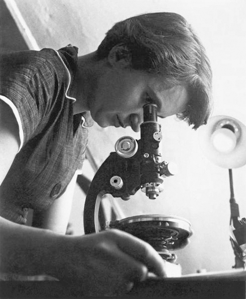
Rosalind Franklin was a British scientist whose research made a major contribution to understanding the structure of DNA. She specialised in X-ray crystallography, a technique used to study the arrangement of atoms in molecules by examining how X-rays scatter when directed at crystals.
In the early 1950s, Franklin worked at King’s College London alongside scientist Maurice Wilkins. Franklin produced detailed X-ray diffraction images of DNA, including the famous Photo 51, which provided evidence of DNA’s helical (spiral) structure.
At the same time, scientists James Watson and Francis Crick at the University of Cambridge were attempting to determine the structure of DNA. Using information from Franklin’s work, along with other scientific evidence, Watson and Crick proposed the double helix model of DNA in 1953.
Franklin’s careful experimental evidence was essential in confirming the dimensions and shape of the DNA molecule, however, her contribution was not fully recognised during her lifetime. In 1962, Watson, Crick and Wilkins received the Nobel Prize in Physiology or Medicine for the discovery of DNA’s structure. Franklin had died in 1958 from ovarian cancer, and Nobel Prizes are not awarded posthumously.
Today, Franklin is recognised as a key contributor to the understanding of the structure of DNA.
References
Cobb, M., & Comfort, N. (2023). What Rosalind Franklin Truly Contributed to the Discovery of DNA’s Structure. Nature, 616(7958), 657–660. https://doi.org/10.1038/d41586-023-01313-5
Rosalind Franklin was a British scientist whose research made a major contribution to understanding the structure of DNA. She specialised in X-ray crystallography, a technique used to study the arrangement of atoms in molecules by examining how X-rays scatter when directed at crystals.
In the early 1950s, Franklin worked at King’s College London alongside scientist Maurice Wilkins. Franklin produced detailed X-ray diffraction images of DNA, including the famous Photo 51, which provided evidence of DNA’s helical (spiral) structure.
At the same time, scientists James Watson and Francis Crick at the University of Cambridge were attempting to determine the structure of DNA. Using information from Franklin’s work, along with other scientific evidence, Watson and Crick proposed the double helix model of DNA in 1953.
Franklin’s careful experimental evidence was essential in confirming the dimensions and shape of the DNA molecule, however, her contribution was not fully recognised during her lifetime. In 1962, Watson, Crick and Wilkins received the Nobel Prize in Physiology or Medicine for the discovery of DNA’s structure. Franklin had died in 1958 from ovarian cancer, and Nobel Prizes are not awarded posthumously.
Today, Franklin is recognised as a key contributor to the understanding of the structure of DNA.
References
Cobb, M., & Comfort, N. (2023). What Rosalind Franklin Truly Contributed to the Discovery of DNA’s Structure. Nature, 616(7958), 657–660. https://doi.org/10.1038/d41586-023-01313-5